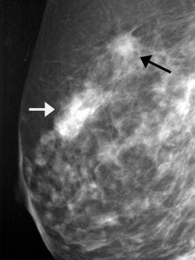
A 60-year-old patient presented by a lump in the left breast
$ 19.99 · 4.8 (679) · In stock

Download scientific diagram | A 60-year-old patient presented by a lump in the left breast. Mammography revealed focal asymmetry in the left upper inner quadrant with microcalcifications (a, b). DBT showed left breast spiculated mass with microcalcifications as well as right breast retroareolar nodule with microcalcifications (c, d). CEM showed left breast heterogeneously enhancing upper inner quadrant mass lesion with spiculated margins and surrounding multiple satellite lesions as well as right breast tiny right retroareolar homogenously enhancing mass with not circumscribed irregular margins (e, f). Breast ultrasound showed left breast irregular ill-defined mass in the left upper inner quadrant as well as right retroareolar small irregular ill-defined mass (g, h). The final diagnosis was bilateral invasive duct carcinoma from publication: Comparative study between contrast-enhanced mammography, tomosynthesis, and breast ultrasound as complementary techniques to mammography in dense breast parenchyma | Background Mammography is accused of having low sensitivity and specificity in dense breast parenchyma. Also, women with dense breasts show an increased risk of developing breast cancer. Breast ultrasound has been used for several years for a better characterization of breast | Breast Ultrasound, Mammography and breast | ResearchGate, the professional network for scientists.

A 60-year-old patient presented by a lump in the left breast.
Breast Cancer Ultrasonography: Practice Essentials, Role of Ultrasonography in Screening, Breast Imaging Reporting and Data System
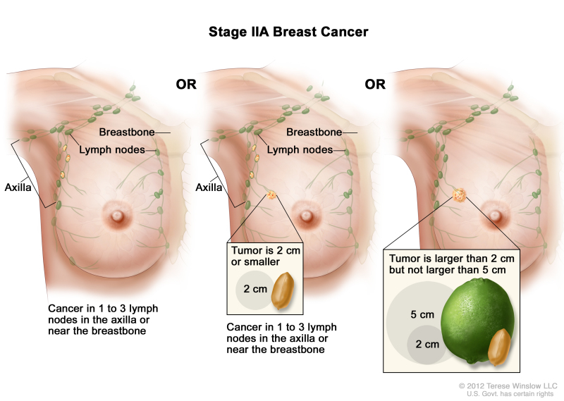
Shorter Radiation Course for Some with Early Breast Cancer - NCI

A Lump Under Your Breast at Bra Line: What to Know

A 60-year-old patient presented by a lump in the left breast.

Performance of AI-aided mammography in breast cancer diagnosis: Does breast density matter?, Egyptian Journal of Radiology and Nuclear Medicine

Rasha Mohamed Kamal's research works Cairo University, Cairo (CU) and other places

J. Imaging, Free Full-Text

A 48-year-old woman complains of a left breast lump. a Craniocaudal and

Breast Cancer Lumps - How to Know if a Lump is Breast Cancer?

PDF) Comparative study between contrast-enhanced mammography, tomosynthesis, and breast ultrasound as complementary techniques to mammography in dense breast parenchyma

A 36-year-old woman complains of a right breast lump. a Craniocaudal